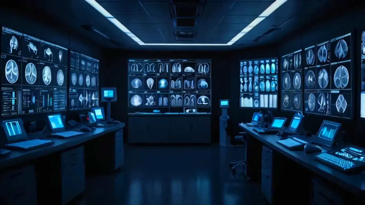
Gestión eficiente de estudios de rayos X en alto volumen
La gestión de estudios de rayos X en centros con alta demanda representa uno de los mayores ...
Optimiza tu flujo de trabajo en clínicas, hospitales y centros radiológicos

Una abogada experta y un médico radiólogo analizan, sin filtros, los vacíos legales que hoy enfrenta el uso de la IA en la radiología.


El problema no es el volumen. Es la ineficiencia oculta.
Te enseñamos a recuperar hasta 2 horas diarias por radiólogo.
Ven al próximo webinar gratuito:
¿Radiología en colapso? Detén las fugas invisibles


Cuando el sistema falla, la operación se resiente: retrasos, reprocesos y pérdida de referidores. Descubre en este webinar cómo fortalecer la relación con tu tecnología y llevar tu servicio de radiología al siguiente nivel.


.webp)
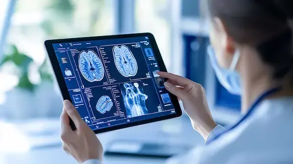
Aquila+ mejora significativamente la productividad clínica, la precisión diagnóstica y la experiencia del radiólogo, más allá de la lectura remota.
.webp)
Simplifica la gestión en los centros radiológicos y el diagnóstico en todos los niveles de complejidad.
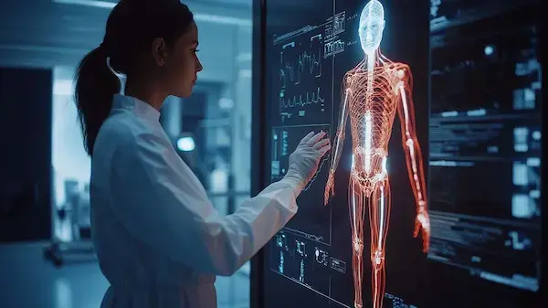
Con algoritmos que contribuyen a interpretar los estudios de radiología de forma más exacta y en menor tiempo.
.png)


.webp)
.webp)
.webp)
.webp)
.webp)
.webp)
.webp)
.webp)
.webp)
.webp)
.webp)
.webp)
.webp)
.webp)

.webp)
.webp)
.webp)
.webp)
.webp)
%20(1).webp)
IMEXHS proporciono experiencia y cumplimiento para los ...
.webp)
IMEXHS realizó el reemplazo de la tecnología por el 60% ...
.webp)
Para nosotros IMEXHS representa innovación, tecnología, y sobre todo nos permite ser muy eficientes y rápidos con todos nuestros pacientes y usuarios.
.webp)
En Latinoamérica la necesidad creciente de una solución radiológica que se pueda adaptar a las posibilidades que existen alrededor y además tener un PACS de gran calidad es sin duda lo que marca a IMEXHS como empresa.

“No es solo un sistema que funciona, es una plataforma que ordena el proceso, reduce fricciones y permite a la administración concentrarse en lo verdaderamente estratégico”.